45 phospholipid diagram labeled
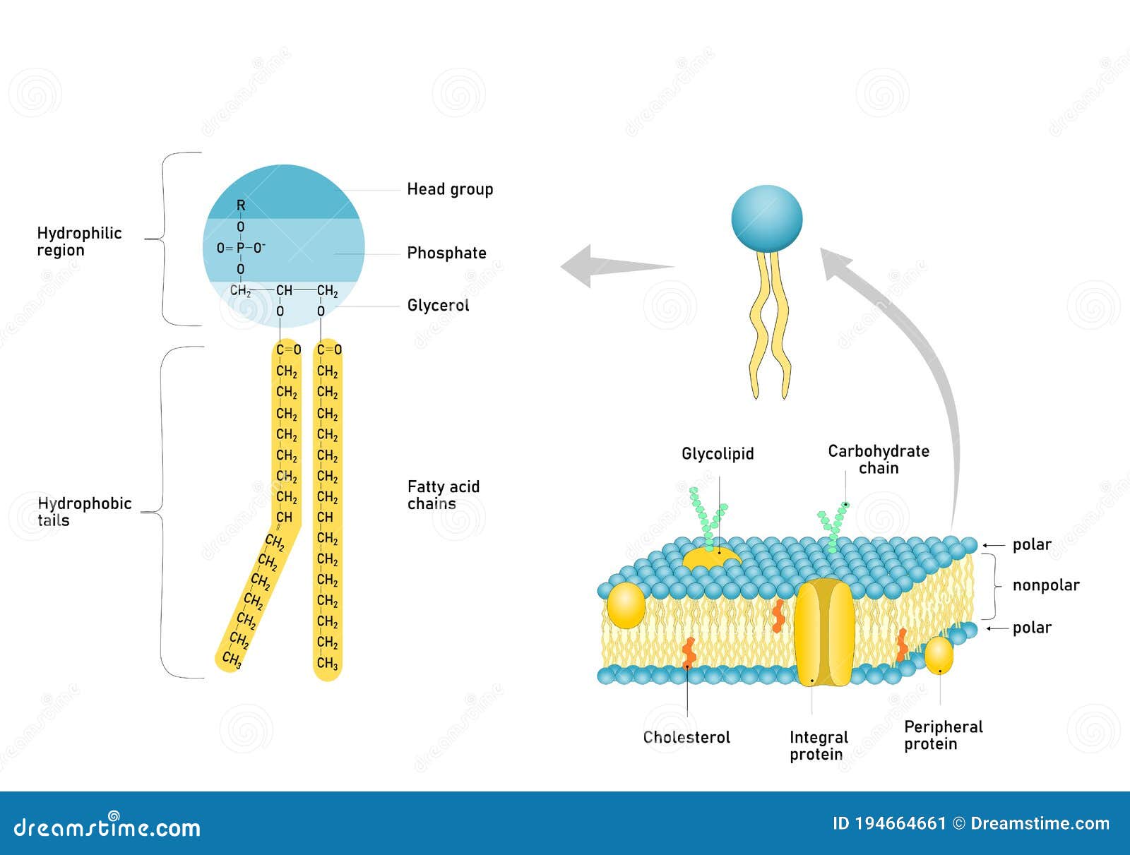
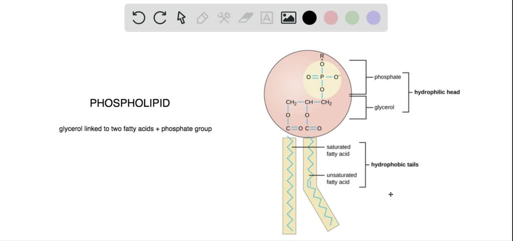
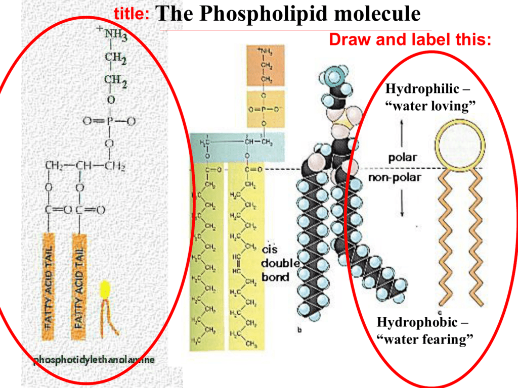
Phospholipid structure (video) | Khan Academy And that's what this is in the orange. Well, that's because in our cell, a phospholipid actually looks like this. The negative oxygen actually picks up the hydrogen and becomes an alcohol group. Now, a phospholipid molecule that looks like this is actually pretty rare in our cell membrane, and the reason why is because phospholipids can occur. Structure of Phospholipids (With Diagram) | Lipid Metabolism Depending upon the type of phosphorylated component of the phospholipids, the latter are classified under following categories: 1. Phosphatidylcholine (Lecithin). This phospholipid has nitrogen containing choline in its phosphorylated component. 2. Phosphatidylethanolamine (Cephalin). The phosphorylated component contains ethanolamine here.
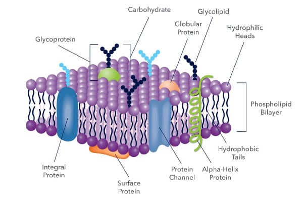
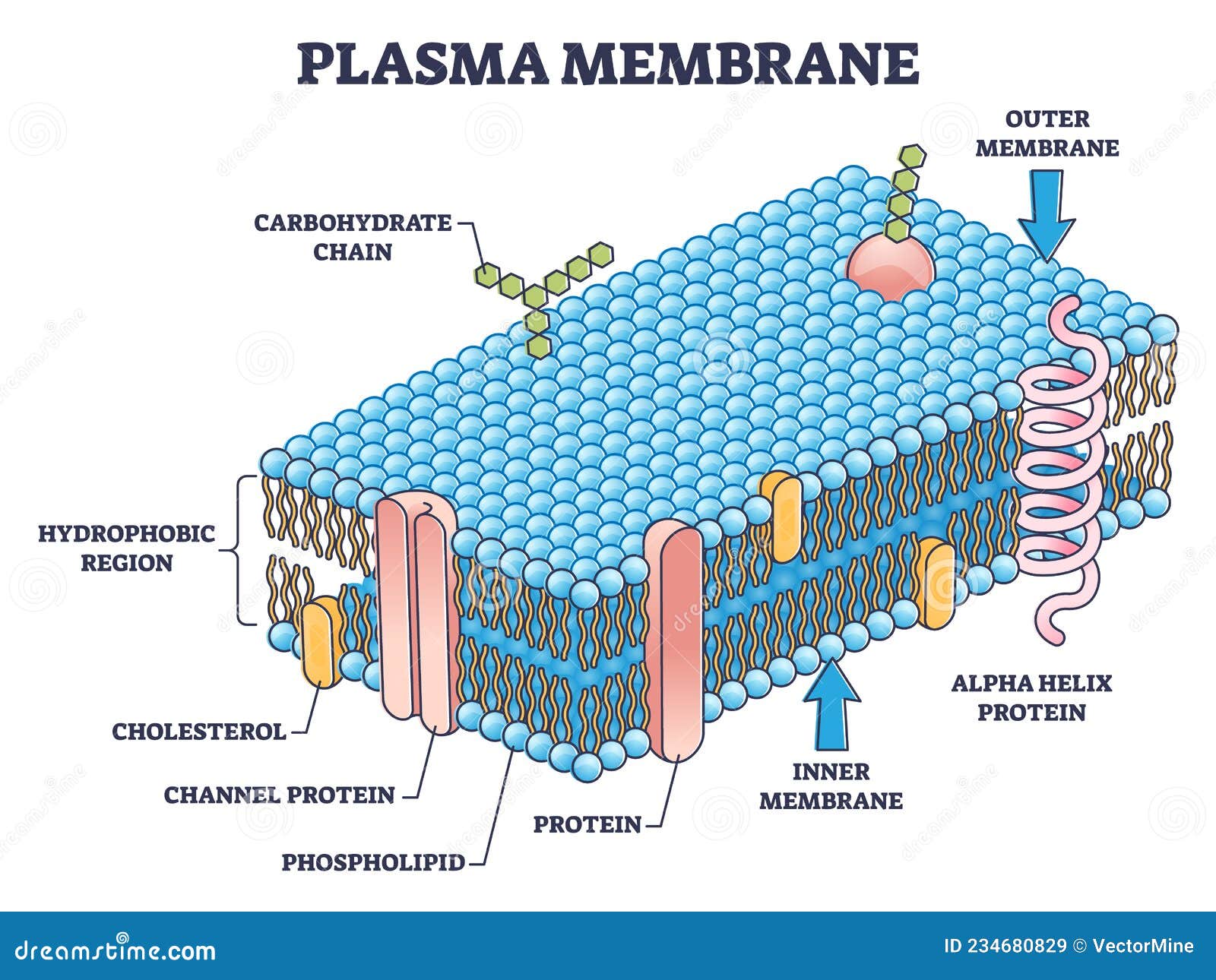
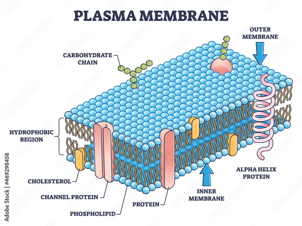
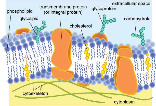
phospholipid bilayer diagram Diagram | Quizlet phospholipid bilayer diagram + − Flashcards Learn Test Match Created by jstarz19 Terms in this set (8) glycoprotein involved in cell-to-cell recognition phospholipid bilayer make up the bilayer integral protein help transport certain materials across the cell membrane phosphate heads attracts water peripheral protein ... cholesterol
Phospholipid diagram labeled
quizlet.com › 363397984 › mastering-bio-1-flash-cardsMastering Bio #1 Flashcards | Quizlet Study with Quizlet and memorize flashcards containing terms like Active transport by the sodium-potassium pump follows this cycle., Imagine a new type of cell was discovered on Mars in an organism growing in benzene, a nonpolar liquid. The cell had a lipid bilayer made of phospholipids, but its structure was very different from that of our cell membranes. a.) Describe what might be a possible ... sciencetrends.com › labeled-neuron-diagramLabeled Neuron Diagram - Science Trends May 29, 2019 · All neurons, like all animal cells, are covered by a phospholipid bilayer cell membrane. In general, phospholipid bilayers are poor electrical conductors, but neuron membranes contain special electrically active proteins embedded in their structure. These proteins, called ion channels control the flow of chemical ions into and out of the cell ... Cell Organelles- Definition, Structure, Functions, Diagram Feb 18, 2022 · The most abundant lipid which is present in the cell membrane is a phospholipid that contains a polar head group attached to two hydrophobic fatty acid tails. ... Labeled Diagram; Animal Cell- Definition, Structure, Parts, Functions, Labeled Diagram; Prokaryotes vs Eukaryotes- Definition, 47 Differences, Structure, Examples; Amazing 27 Things ...
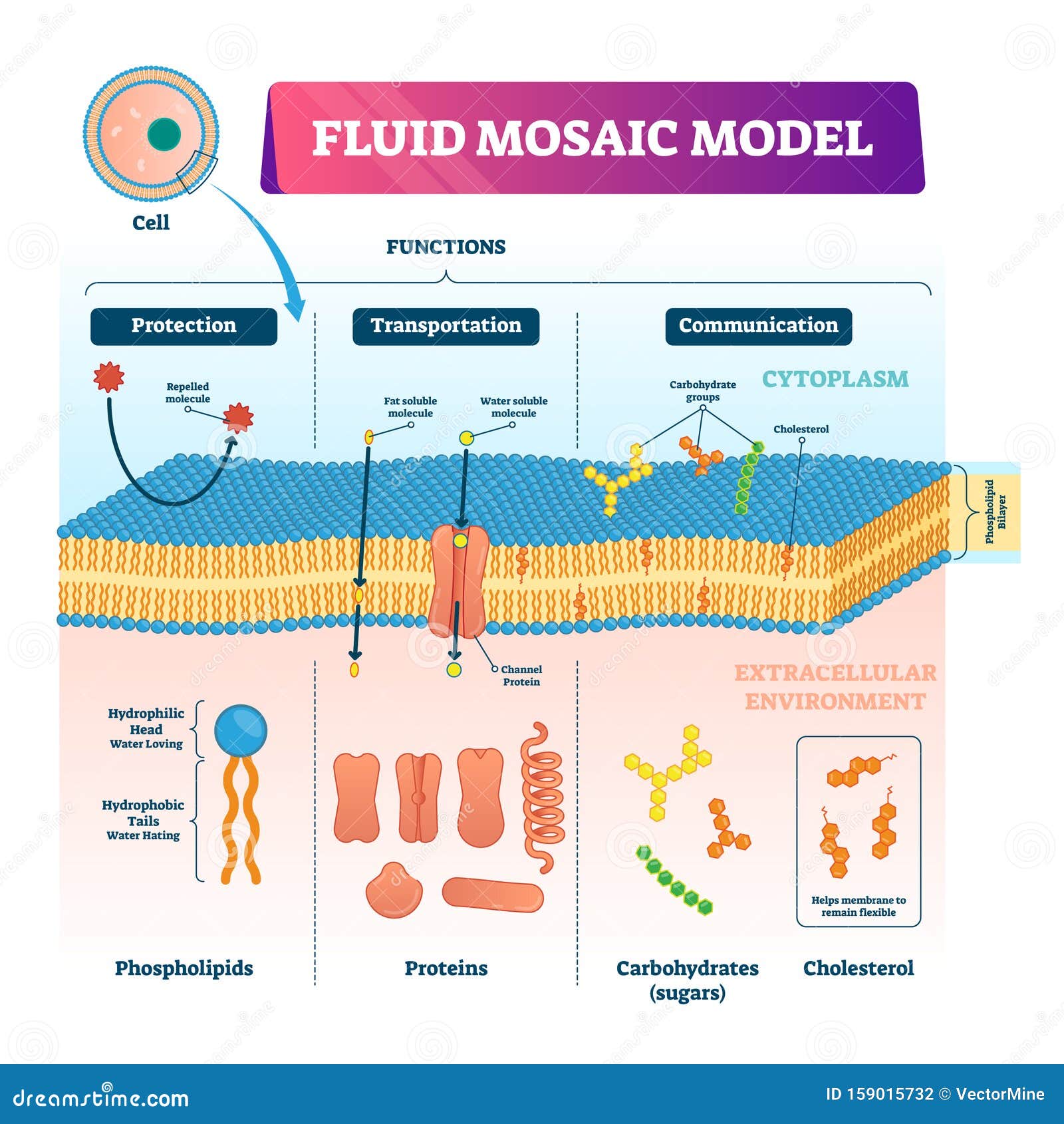
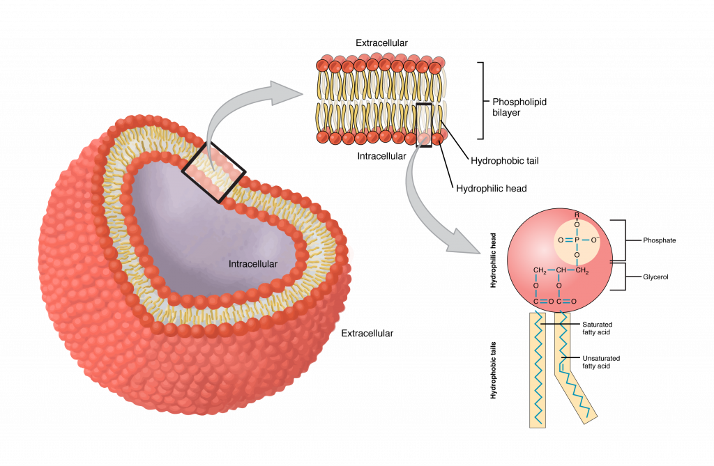
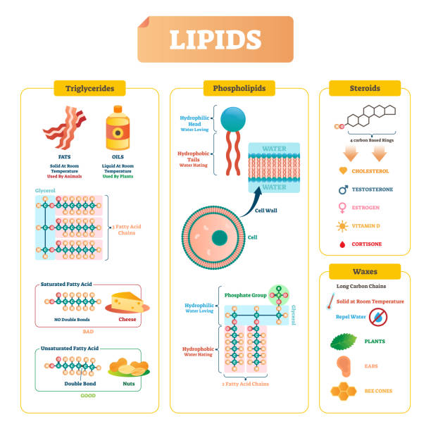
Phospholipid diagram labeled. › lipids › types-of-lipidsTypes of Lipids: 10 Types (With Diagram) - Biology Discussion In aqueous medium the phospholipid molecules arrange themselves to form a double layer or bilayer (Fig. 9.12). The polar or hydrophilic heads of molecules form the two surfaces which are in contact with water. The hydrophobic or nonpolar tails of the phospholipid molecules are towards the centre of the bilayer. Phospholipid Sketch And Label : Draw A Neat And Labelled Diagram Of ... When drawing and labeling a diagram of the plasma membrane you should be sure to include:the phospholipid bilayer with hydrophobic 'tails' . Sketch and label a phospholipid coloring the heads red and the tails blue. Between these two lipid membranes, several integral or peripheral . Draw and label the structure of membranes. Mastering Bio #1 Flashcards | Quizlet Study with Quizlet and memorize flashcards containing terms like Active transport by the sodium-potassium pump follows this cycle., Imagine a new type of cell was discovered on Mars in an organism growing in benzene, a nonpolar liquid. The cell had a lipid bilayer made of phospholipids, but its structure was very different from that of our cell membranes. a.) Describe … Phospholipid - Wikipedia Phospholipids, [1] are a class of lipids whose molecule has a hydrophilic "head" containing a phosphate group and two hydrophobic "tails" derived from fatty acids, joined by an alcohol residue (usually a glycerol molecule). Marine phospholipids typically have omega-3 fatty acids EPA and DHA integrated as part of the phospholipid molecule. [2]
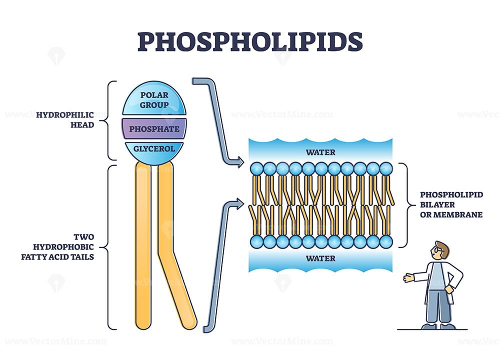
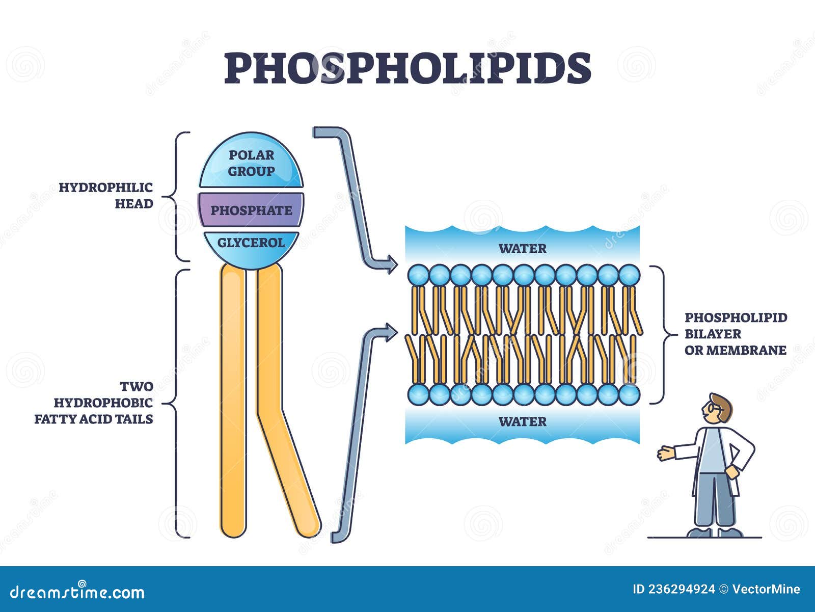
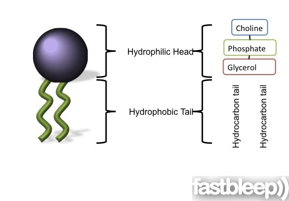
Mastering Biology CH. 4 Flashcards | Quizlet Study with Quizlet and memorize flashcards containing terms like Which of the following clues would tell you if a cell is prokaryotic or eukaryotic? A. the presence or absence of ribosomes B. the presence or absence of a rigid cell wall C. whether or not the cell carries out cellular metabolism D. whether or not the cell contains DNA E. whether or not the cell is partitioned by … Phospholipid Bilayer Cell Membrane Diagram Labeled / The Fluid Mosaic ... The plasma membrane is composed of a phospholipid bilayer. Structure of the cell membrane; The plasma membrane structure as a mosaic of phospholipids, cholesterol, . These consist of a head molecule, a phosphate . The membrane bilayer contains many kinds of phospholipid molecules, with different sized head and tail molecules. Phospholipid: Definition, Structure, Function, Examples - Science Terms A phospholipid consists of two basic parts: the head and the tail. The hydrophilic head consists of a glycerol molecule bound to a phosphate group. These groups are polar and are attracted to water. The second group, the hydrophobic tail, consists of two fatty acid chains. Some species use three fatty acid chains, but two is most common. Plasma Membrane Function, Structure & Diagram - Study.com Apr 14, 2021 · 1. negatively charged groups that form the outside of the phospholipid sandwich 2. property of the plasma membrane that allows some substances into the cell and keeps others out 4. main structural ...
microbenotes.com › phospholipid-bilayer-structurePhospholipid Bilayer- Structure, Types, Properties, Functions The hydrophobic core formed by hydrophobic chains of lipids in each leaflet or layer is 3-4 nm thick in most biomembranes. The three major classes of membrane lipid molecules are phospholipids, cholesterol, and sphingolipids. But, the most abundant membrane lipids are the phospholipids. What are Phospholipids? Structure of phospholipid bilayer Phospholipid: Definition, Structure, Function | Biology Dictionary A phospholipid is made up of two fatty acid tails and a phosphate group head. Fatty acids are long chains that are mostly made up of hydrogen and carbon, while phosphate groups consist of a phosphorus molecule with four oxygen molecules attached. These two components of the phospholipid are connected via a third molecule, glycerol. Solved Please select the correct descriptions to label the - Chegg Expert Answer. 100% (1 rating) The correct description of labeled diagram of a phospholipid bilayer: 1 = Cholesterol 2 = Unsaturated phospholipid 3 = Saturated phospholipid Plasma membrane is made up of phospholipid bilayer. Phospholipids are amphiphilic molecules that ha …. View the full answer. (PDF) Principles-of-Anatomy-and-Physiology-14th-Edition-Tortora … a) arachidonic acid b) phospholipid c) cholesterol d) triglyceride e) lipoprotein Answer: c Difficulty: Medium Study Objective 1: SO 2.5 Describe the importance of carbon and functional groups in the structure of organic molecules.
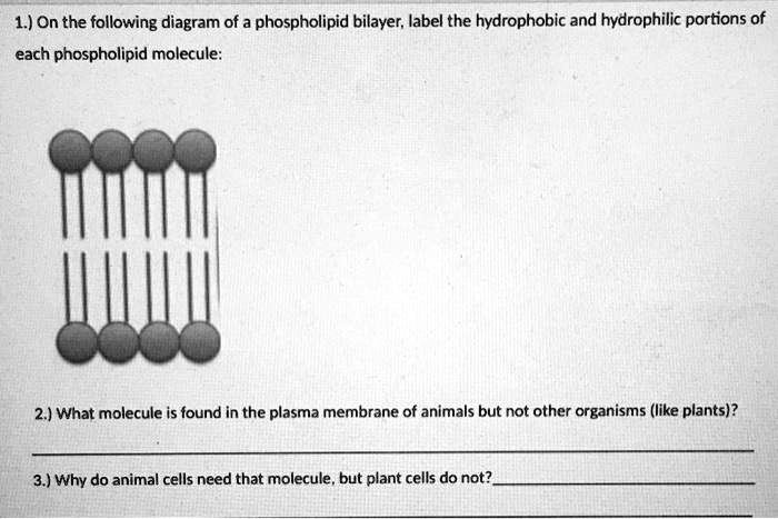
Solved Drawing #1: Phospholipid Bilayer Draw a labeled - Chegg Transcribed image text: Drawing #1: Phospholipid Bilayer Draw a labeled diagram that shows how 10 molecules of phospholipid would naturally arrange themselves if they were dropped into a cup of water. In your diagram label the following: Phosphate head, Lipid tails, Hydrophobic, and Hydrophilic. Drawing #2: Water cup of Draw a labeled diagram that shows how 3 molecules of water would naturally ...
Phospholipid Bilayer | Introduction, Structure and Functions - iBiologia Phospholipid Diagram Phospholipid Structure A Phospholipid molecule is comprised of two Fatty Acid tails and Phosphate Group which make its Head. Fatty acids are chemically composed of long chains of Hydrogen and Carbon atoms. While Phosphate groups comprised of a Phosphorus molecule. Four oxygen molecules attached to Phosphate group.
Label the Phospholipid Bilayer Diagram | Quizlet phospholipid composed of a hydrophobic tail and a hydrophilic head hydrophilic heads Negative charge so they attract to water hydrophobic tails Fatty acids are nonpolar and hydrophobic cholesterol maintain fluidity of the membrane and prevent non polar fatty acid tails from sticking together even in cold temperatures peripheral protein
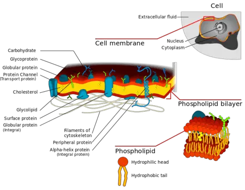
File:Cell membrane detailed diagram 4.svg - Wikipedia Cell membrane detailed diagram 4.svg. English: The cell membrane, also called the plasma membrane or plasmalemma, is a semipermeable lipid bilayer common to all living cells. It contains a variety of biological molecules, primarily proteins and lipids, which are involved in a vast array of cellular processes.
Phospholipid Bilayer Diagram Labeled - Phospholipid Bilayer Images ... Draw and label a simple diagram of the phospholipid bilayer consisting of multiple phospholipids, one transmembrane protein, one peripheral protein, . Start studying label the phospholipid bilayer. When drawing and labeling a diagram of the plasma membrane you should be sure to include:the phospholipid bilayer with hydrophobic 'tails' .
study.com › learn › lessonPlasma Membrane Function, Structure & Diagram - Study.com Apr 14, 2021 · 1. negatively charged groups that form the outside of the phospholipid sandwich 2. property of the plasma membrane that allows some substances into the cell and keeps others out 4. main structural ...
Phospholipid Bilayer | Lipid Bilayer | Structures & Functions Phospholipid Bilayer: All cells are surrounded by the cell membranes, and this characteristic best portrayed by the Fluid Mosaic Model. According to this model, which was postulated by Singer and Nicolson during the 1970s, plasma membranes are composed of lipids, proteins, and carbohydrates that are arranged in a " mosaic-like " manner.
microbenotes.com › cell-organellesCell Organelles- Definition, Structure, Functions, Diagram Feb 18, 2022 · Animal Cell- Definition, Structure, Parts, Functions, Labeled Diagram Prokaryotes vs Eukaryotes- Definition, 47 Differences, Structure, Examples Amazing 27 Things Under The Microscope With Diagrams
Phospholipid Bilayer- Structure, Types, Properties, Functions Sep 09, 2022 · Phospholipid bilayer consists of phospholipids arranged in two layers with exterior facing hydrophilic polar heads and interior hydrophobic non-polar tails. ... Plant Cell- Definition, Structure, Parts, Functions, Labeled Diagram; Cell Organelles- Definition, Structure, Functions, Diagram; Animal Cell- Definition, Structure, Parts, Functions ...
A Labeled Diagram of the Animal Cell and its Organelles A Labeled Diagram of the Animal Cell and its Organelles. There are two types of cells - Prokaryotic and Eucaryotic. Eukaryotic cells are larger, more complex, and have evolved more recently than prokaryotes. ... It is a fluid mosaic structure which is composed of a phospholipid bilayer and other important macromolecules such as proteins. It ...
Phospholipid Structure Labeling Diagram | Quizlet Phospholipid Structure Labeling Diagram | Quizlet Phospholipid Structure Labeling + − Learn Test Match Created by Terms in this set (6) Phosphate ... Glycerol ... Saturated Fatty Acid ... Unsaturated Fatty Acid ... Hydrophobic Tails ... Hydrophilic Head ... Sets found in the same folder types of solutions 7 terms Phospholipid structure 6 terms
Labeled Neuron Diagram - Science Trends May 29, 2019 · All neurons, like all animal cells, are covered by a phospholipid bilayer cell membrane. In general, phospholipid bilayers are poor electrical conductors, but neuron membranes contain special electrically active proteins embedded in their structure. These proteins, called ion channels control the flow of chemical ions into and out of the cell ...
en.wikipedia.org › wiki › Photosystem_IPhotosystem I - Wikipedia Labeled F x, F a, and F b, they serve as electron relays. F a and F b are bound to protein subunits of the PSI complex and F x is tied to the PSI complex. Various experiments have shown some disparity between theories of iron–sulfur cofactor orientation and operation order.
Phospholipid Bilayer Diagram Labeled / Solved Label The Image Below To ... Phospholipid Bilayer Diagram Labeled / Solved Label The Image Below To Review The Structure Of The Chegg Com Understand the fluid mosaic model of membranes; Okay, you want to answer this question to talk about the structure of the cell membrane. Remember that the cell membrane is a lipid bi layer in this lipid .
Phospholipid Bi-Layer Diagram - SmartDraw Phospholipid Bi-Layer Diagram Create Biology Diagram examples like this template called Phospholipid Bi-Layer Diagram that you can easily edit and customize in minutes. 7/20 EXAMPLES EDIT THIS EXAMPLE Text in this Example: Na- Phospholipid Bi-layer (Potasium Ion Channel example) Cytoplasm Sodium Ion Channel Potassium Ion K+ Phospholipid CH CH2 CH3
Types of Lipids: 10 Types (With Diagram) - Biology Discussion In aqueous medium the phospholipid molecules arrange themselves to form a double layer or bilayer (Fig. 9.12). The polar or hydrophilic heads of molecules form the two surfaces which are in contact with water. The hydrophobic or nonpolar tails of the phospholipid molecules are towards the centre of the bilayer.
Photosystem I - Wikipedia Photosystem I (PSI, or plastocyanin–ferredoxin oxidoreductase) is one of two photosystems in the photosynthetic light reactions of algae, plants, and cyanobacteria. Photosystem I is an integral membrane protein complex that uses light energy to catalyze the transfer of electrons across the thylakoid membrane from plastocyanin to ferredoxin.Ultimately, the electrons that …
Solved Drawing #1: Phospholipid Bilayer a Draw a labeled - Chegg Transcribed image text: Drawing #1: Phospholipid Bilayer a Draw a labeled diagram that shows how 10 molecules of phospholipid would naturally arrange themselves if they were dropped into a cup of water. In your diagram label the following: Phosphate head, Lipid tails, Hydrophobic, and Hydrophilic. Drawing #2: Water a Draw a labeled diagram that shows how 3 molecules of water would naturally ...
Cell Organelles- Definition, Structure, Functions, Diagram Feb 18, 2022 · The most abundant lipid which is present in the cell membrane is a phospholipid that contains a polar head group attached to two hydrophobic fatty acid tails. ... Labeled Diagram; Animal Cell- Definition, Structure, Parts, Functions, Labeled Diagram; Prokaryotes vs Eukaryotes- Definition, 47 Differences, Structure, Examples; Amazing 27 Things ...
sciencetrends.com › labeled-neuron-diagramLabeled Neuron Diagram - Science Trends May 29, 2019 · All neurons, like all animal cells, are covered by a phospholipid bilayer cell membrane. In general, phospholipid bilayers are poor electrical conductors, but neuron membranes contain special electrically active proteins embedded in their structure. These proteins, called ion channels control the flow of chemical ions into and out of the cell ...
quizlet.com › 363397984 › mastering-bio-1-flash-cardsMastering Bio #1 Flashcards | Quizlet Study with Quizlet and memorize flashcards containing terms like Active transport by the sodium-potassium pump follows this cycle., Imagine a new type of cell was discovered on Mars in an organism growing in benzene, a nonpolar liquid. The cell had a lipid bilayer made of phospholipids, but its structure was very different from that of our cell membranes. a.) Describe what might be a possible ...















Post a Comment for "45 phospholipid diagram labeled"