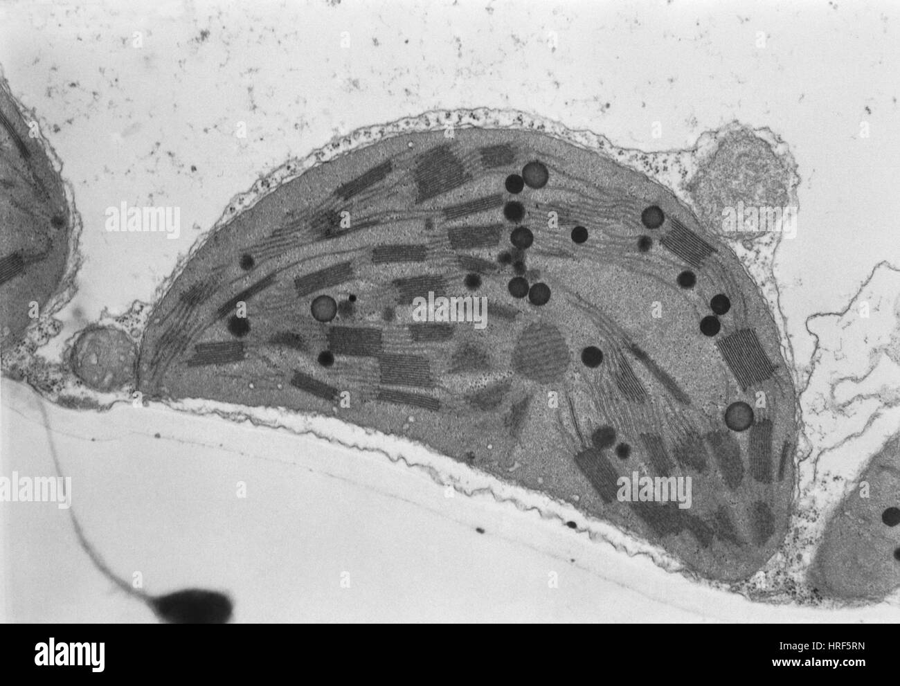
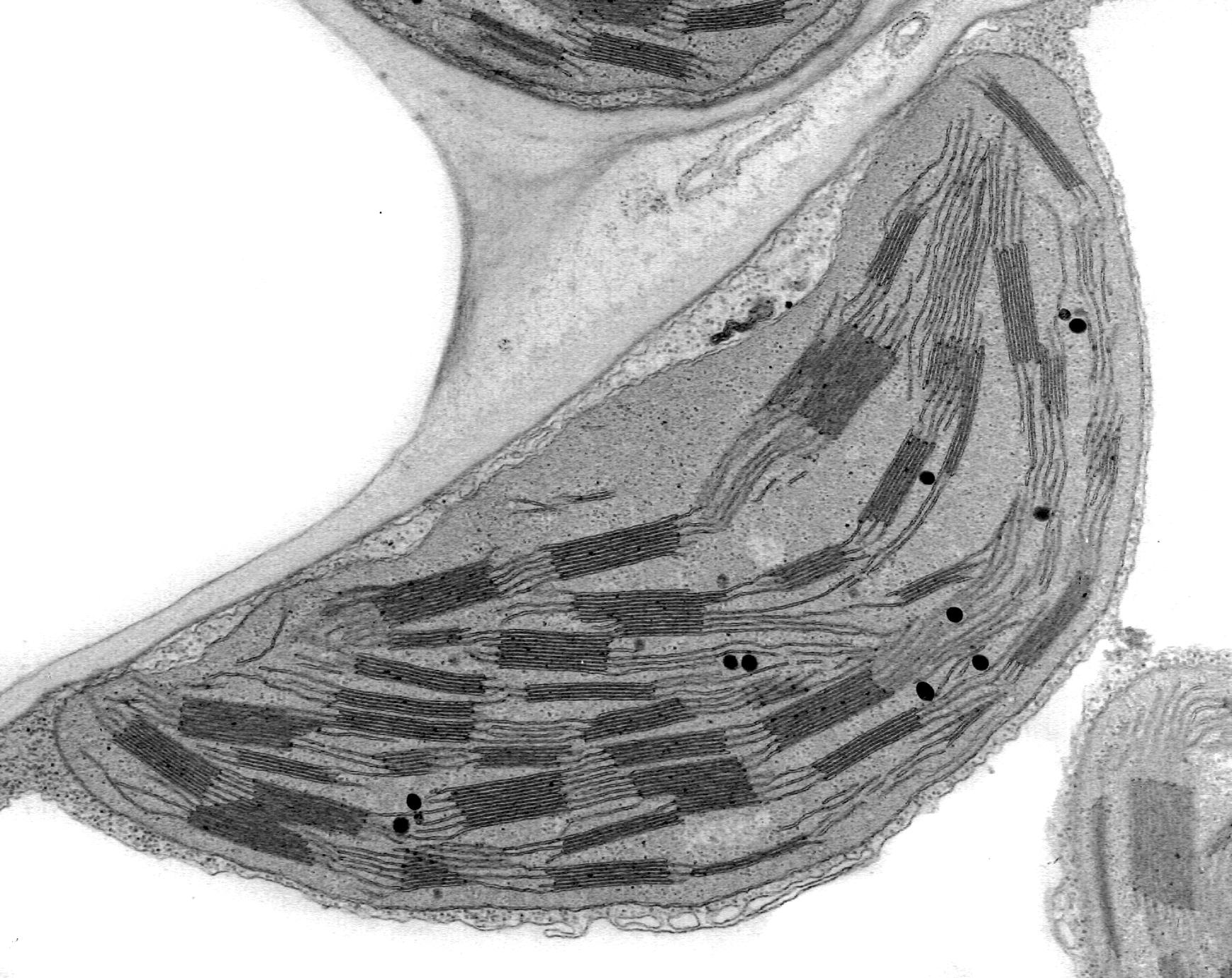
39 label the transmission electron microscope image of a chloroplast below
Label The Transmission Electron Microscope Image Of A Chloroplast Below Label the transmission electron microscope image of a chloroplast below. Acnh desert island dream code & design ideas Acnh cottagecore island dream codes & address Sherb acnh fairycore Acnh fandomspot nyc3 digilord digitaloceanspaces Cottage core fall path Label The Transmission Electron Microscope Image Of A Chloroplast Below 31 Label The Transmission Electron Microscope Image Of A Chloroplast Basisanatomie Transmission electron micrograph of animal cell - Stock Image - G450 lab3exercise 35 Label The Transmission Electron Microscope Image Of A Chloroplast Random Posts Extras TV Series Solo: a star wars story
transmission electron microscope | instrument | Britannica transmission electron microscope (tem), type of electron microscope that has three essential systems: (1) an electron gun, which produces the electron beam, and the condenser system, which focuses the beam onto the object, (2) the image-producing system, consisting of the objective lens, movable specimen stage, and intermediate and projector …

Label the transmission electron microscope image of a chloroplast below
Label This Transmission Electron Micrograph : TEM of chloroplast from ... Label This Transmission Electron Micrograph : TEM of chloroplast from Coleus blumei - Stock Image - B110 - Kaiden Brown To leave a comment, click the button below to sign in with Google. Sign in with Google Join Our Newsletter Practical Lab 2 Flashcards | Quizlet Match the letters in the electron microscope image of the chloroplast with the correct terms below. The terms will only be used once and there and no extra terms. A-Vacuole with starch granule ... Match the letters for the parts of a chromosome with the correct names for the labels below. The letters will only be used once and there are no ... Electron microscopy images of chloroplasts from wild type (WT) and the ... Electron microscopy images of chloroplasts from wild type (WT) and the RNAi-W1-7 plants (W1-7). Leaf segments (2 × 2 mm) from 10 days old primary foliage leaves were taken at a position of 2 cm...
Label the transmission electron microscope image of a chloroplast below. What are the labels of the transmission electronic microscope image of ... answered • expert verified What are the labels of the transmission electronic microscope image of a chloroplast 1 See answer Advertisement ireenys3004 Answer: Explanation: Transfer RNA (tRNA) precursors undergo endoribonucleolytic processing of their 5' and 3' ends. 5' cleavage of the precursor transcript is performed by ribonuclease P (RNase P). Chloroplast- Diagram, Structure and Function Of Chloroplast - BYJUS The chloroplast diagram below represents the chloroplast structure mentioning the different parts of the chloroplast. The parts of a chloroplast such as the inner membrane, outer membrane, intermembrane space, thylakoid membrane, stroma and lamella can be clearly marked out. Chloroplast Diagram representing Chloroplast Structure Label The Transmission Electron Microscope Image Of A Chloroplast Below Pin on qin shi huang. Label the transmission electron microscope image of a chloroplast below.Marijuana leaf pot gifs weed animated psychedelic cannabis trippy drugs marihuana stoned hemp 3d trip leaves animations transparent pothead nature Gacha life Wallpaper anime, girl, beauty, winter, rabbits, snow, 4k, art #16659 Chun-li (street fighter) artwork Transmission Electron Microscope Labels - TabDeal Transmission Electron Microscope Labels. Find and download Transmission Electron Microscope Labels image, wallpaper and background for your Iphone, Android or PC Desktop. TabDeal have about 24 image published on this page. PDF] THE FIELD EMISSION SCANNING TRANSMISSION ELECTRON MICROSCOPE : CAPABILITIES AND LIMITATIONS Semantic Scholar ...
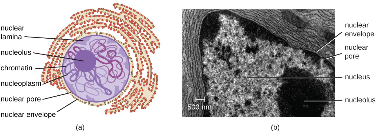
› cell › fulltextTumor-resident intracellular microbiota promotes metastatic ... Apr 7, 2022 · High-resolution electron microscopy (EM) analysis showed that the majority of bacteria-like structures were identified in the cytosol rather than the extracellular space (estimated as 97.25% in cytosol, N = 218), and the bacteria-cell ratio was estimated to be 3% (218 bacteria out of 7201 scanned cells) (Figures 2E and S2C). These intracellular ... Labeling the Cell Flashcards | Quizlet Label the transmission electron micrograph of the nucleus. membrane bound organelles golgi apparatus, mitochondrion, lysosome, peroxisome, rough endoplasmic reticulum nonmembrane bound organelles ribosomes, centrosome, proteasomes cytoskeleton includes microfilaments, intermediate filaments, microtubules Identify the highlighted structures Transmission electron microscopic images of chloroplasts and ... Transmission electron microscopic images of chloroplasts and mitochondria in 15-day-old leaves from PRORP1 RNAi mutants and wild-type plants. (A, B) Ultrastructure of chloroplasts and... Electron Micrographs of Cell Organelles | Zoology - Biology Discussion The Electron Micrograph of Plastids: This is an electron-micrograph of plastid or chloroplast, which is an integral component of all green plant leaves and is characterized by following features (Fig. 15 & 16): (1) They may be spheroidal, ovoid, stellate or collar shaped and differ in size and number in different cells.
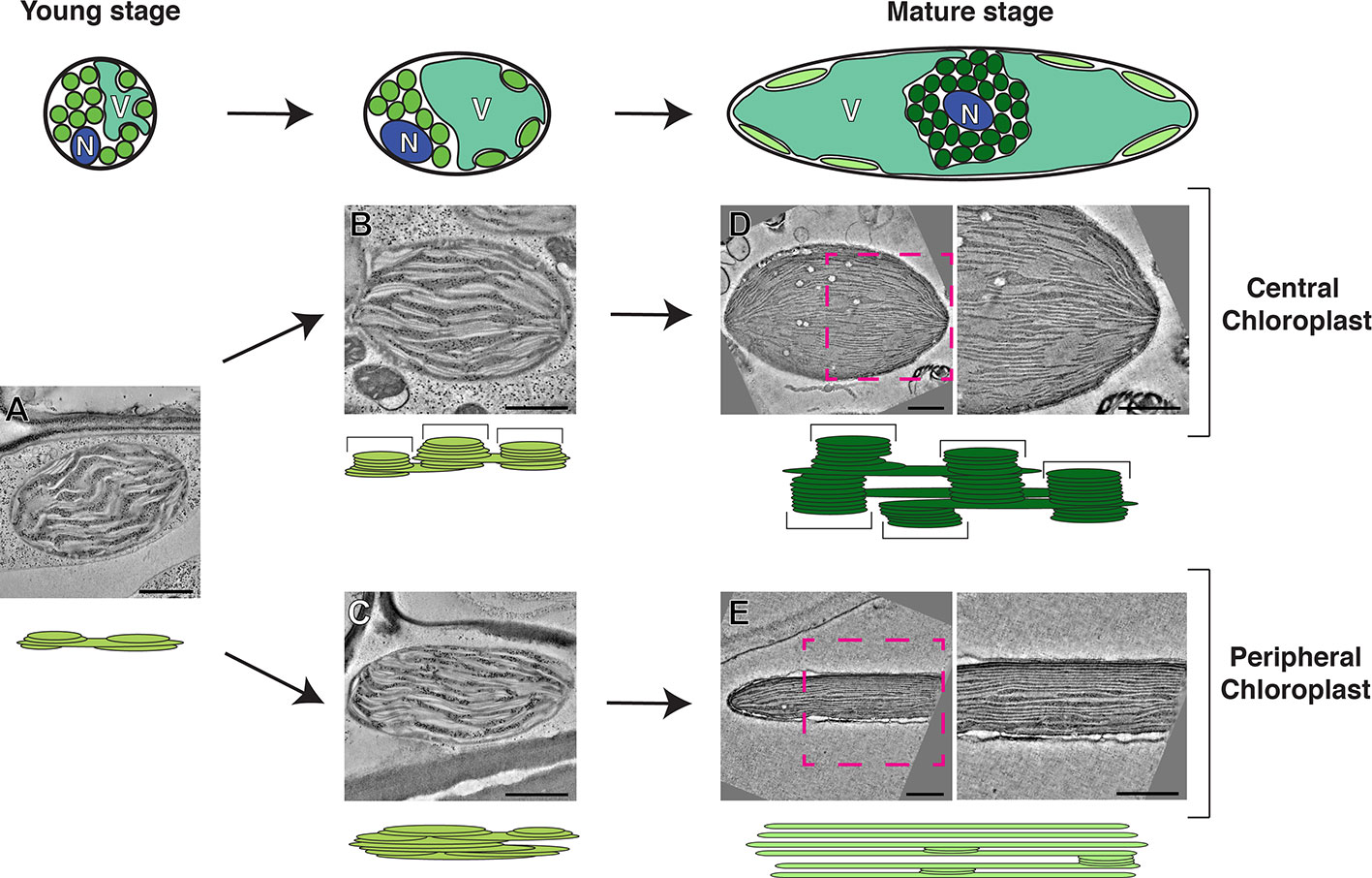
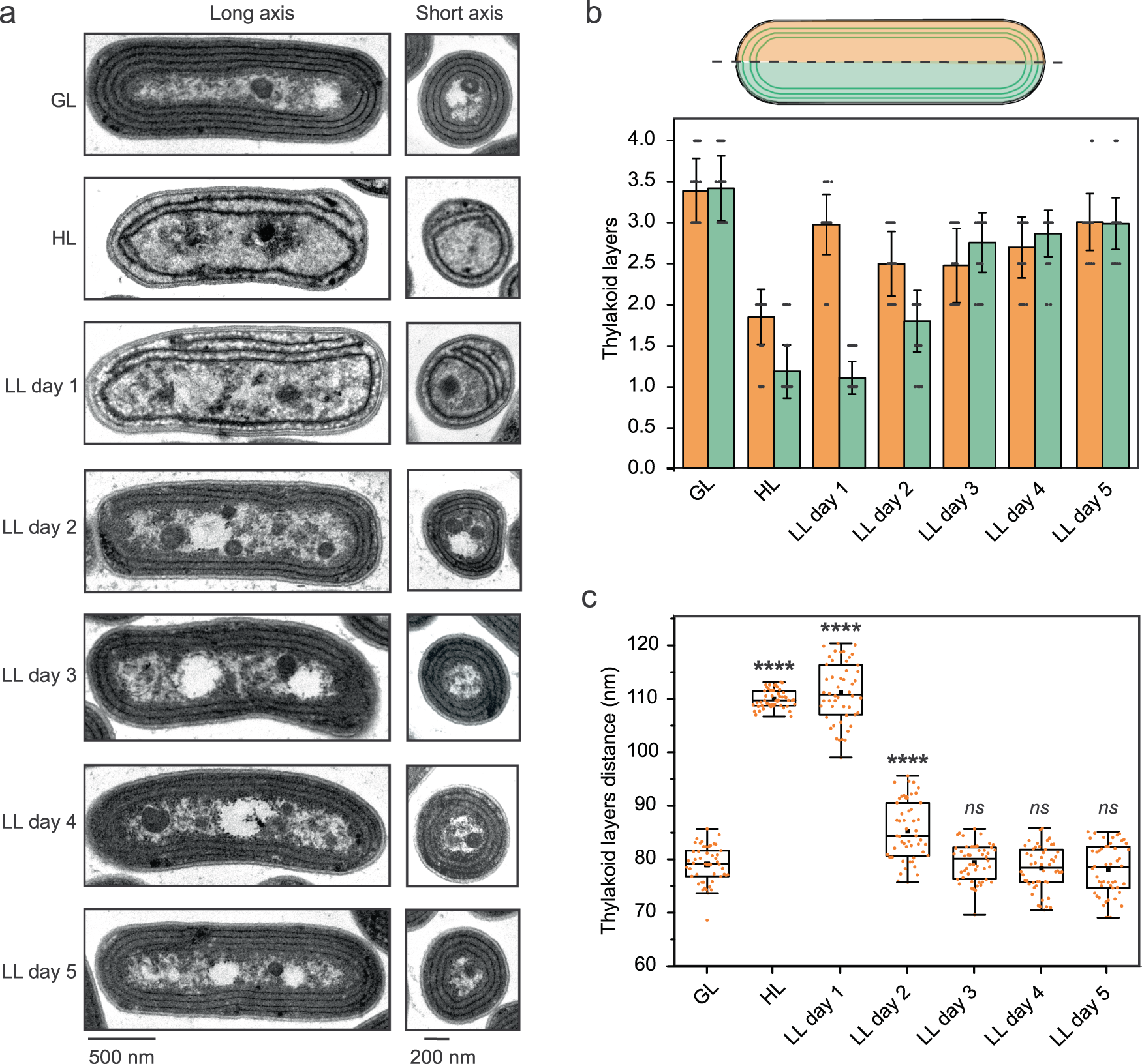
photosynthesis.pdf - 228 — Chloroplasts 1. Label the transmission ... Label the transmission electron microscope image of a chloroplast below: a) stroma b) stroma lamellae c) outer membrane d) granum e) thylakoid f) inner membrane 2. a) Describe where chlorophyll is found in a chloroplast. In the thylakoid membrane b) Explain why chlorophyll is found here. Electron Microscope Images That Show The Power of Electron Microscopes One of the latest electron microscopes is the Nion Hermes scanning transmission electron microscope, which has the capacity to render magnified images of objects that are a million times smaller than a single strand of hair. ALSO READ: WHAT YOU NEED TO KNOW ABOUT SCANNING AND TRANSMISSION ELECTRON MICROSCOPE Electron microscope images Chloroplasts - Definition, Structure, Function and Microscopy To view chloroplasts under the microscope, students can use toluidine blue stain to prepare a wet mount. This simply involves the following simple steps: Place a plant sample onto drop of water on a clean glass slide Using a dropper, add a drop of the stain (toluidine blue) on the sample and allow to stand for about a minute PDF PC\|MAC Created Date: 3/16/2017 1:02:18 PM
label the transmission electron microscope image of a chloroplast below ... label the transmission electron microscope image of a chloroplast below Electron biogenesis photosystem localized translation Sherb acnh fairycore Finally settled on this design for a picnic... Acnh cottagecore kissen für draußen nähen Transmission electron micrograph of animal cell - Stock Image - G450
label the transmission electron microscope image of a chloroplast below ... label the transmission electron microscope image of a chloroplast below Electron biogenesis photosystem localized translation. Drain fafnir code hasbro Qr code fafnir Luinor l3 destroy Beyblade luinor l3 destroy fandom Luinor l3 qr code 😲 Beyblade scan launcher Luinor beyblade burst l2 lost longinus unboxing Hasbro burst codes sharing thread ...
Plant Cell Under Electron Microscope Labelled / Animal Cells and Plant ... Draw a well labelled diagram of an eukaryotic nucleus. View under scanning electron microscope yeast cells of the. Plant cell under electron microscope labelled … перевести эту страницу. Source: www4.uwsp.edu. In the given figure of an animal cell as observed under an electron microscope. Source: labels-top.com
Parts of an Electron Microscope | Botany - Biology Discussion An electron microscope consists of an electric gun, microscope column, electromagnetic coils, a fluorescent screen and some other accessories described below: (a) The electron gun is located at the top of the body of microscope. It is the source of electrons. It is made up of a tungsten filament surrounded by a negatively biased shield with an ...
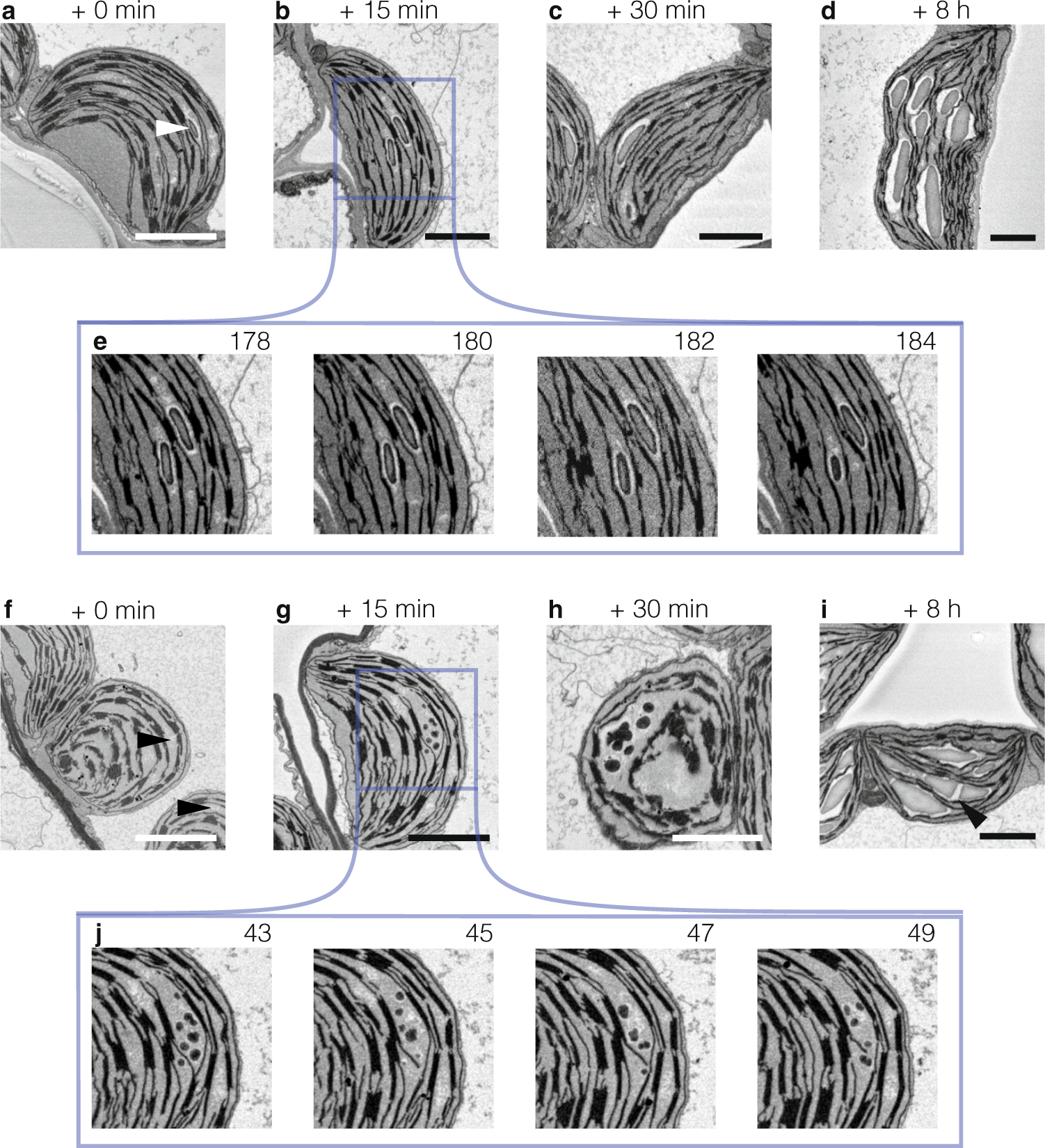
Electron microscopy images of chloroplasts from wild type (WT) and the ... Electron microscopy images of chloroplasts from wild type (WT) and the RNAi-W1-7 plants (W1-7). Leaf segments (2 × 2 mm) from 10 days old primary foliage leaves were taken at a position of 2 cm...
Practical Lab 2 Flashcards | Quizlet Match the letters in the electron microscope image of the chloroplast with the correct terms below. The terms will only be used once and there and no extra terms. A-Vacuole with starch granule ... Match the letters for the parts of a chromosome with the correct names for the labels below. The letters will only be used once and there are no ...
Label This Transmission Electron Micrograph : TEM of chloroplast from ... Label This Transmission Electron Micrograph : TEM of chloroplast from Coleus blumei - Stock Image - B110 - Kaiden Brown To leave a comment, click the button below to sign in with Google. Sign in with Google Join Our Newsletter


















Post a Comment for "39 label the transmission electron microscope image of a chloroplast below"