39 compound microscope diagram
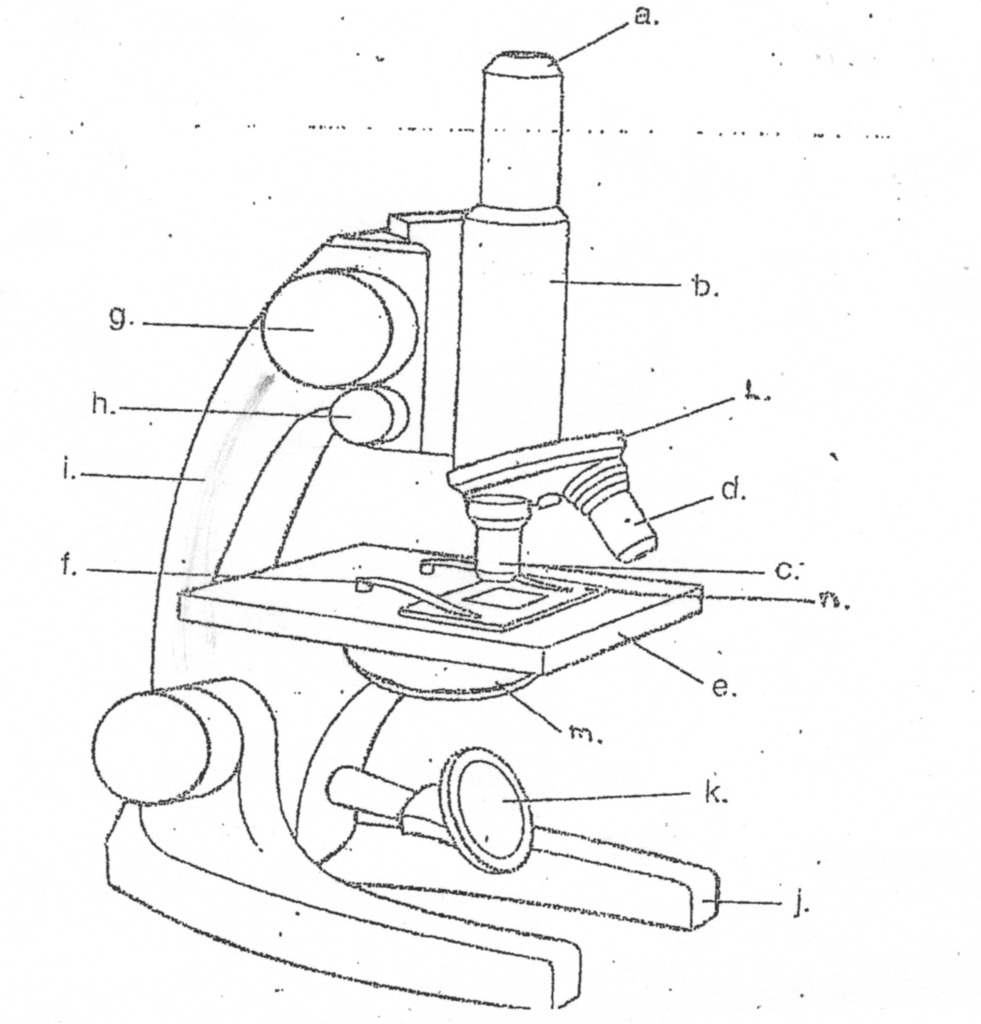
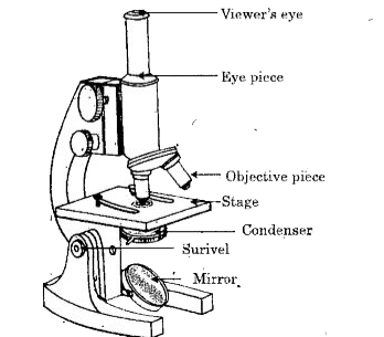
Compound Microscope: Know Definition,working, diagram, properties Compound Microscope Diagram The compound microscope is used to study the structural intricacies of cells, tissues, or organ parts. A compound microscope's components are divided into two categories: Non-optical components Base: The base, often known as the foot, is either U-shaped or horseshoe-shaped. Compound Microscope Parts, Functions, and Labeled Diagram Compound Microscope Definitions for Labels Eyepiece (ocular lens) with or without Pointer: The part that is looked through at the top of the compound microscope. Eyepieces typically have a magnification between 5x & 30x. Monocular or Binocular Head: Structural support that holds & connects the eyepieces to the objective lenses.
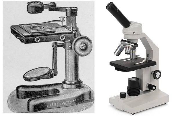
Compound Microscope- Definition, Labeled Diagram, Principle, Parts, Uses Compound microscopes have a combination of lenses that enhances both magnifying powers as well as the resolving power. The specimen or object, to be examined is usually mounted on a transparent glass slide and positioned on the specimen stage between the condenser lens and objective lens.

Compound microscope diagram
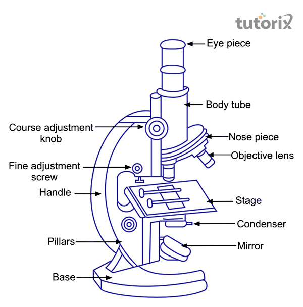
Compound Microscope: Parts of Compound Microscope - BYJU'S The parts of the compound microscope can be categorized into: Mechanical parts; Optical parts (A) Mechanical Parts of a Compound Microscope. 1. Foot or base. It is a U-shaped structure and supports the entire weight of the compound microscope. 2. Pillar. It is a vertical projection. This stands by resting on the base and supports the stage. 3. Arm Compound Microscope Parts, Function, & Diagram - Study.com What are the parts of the compound microscope and their functions? There are many parts to the compound microscope. The eyepiece is the piece a person looks through. The objective lenses... Microscopy: Intro to microscopes & how they work (article) - Khan Academy The fancier instruments that we typically think of as microscopes are compound microscopes, meaning that they have multiple lenses. Because of the way these lenses are arranged, they can bend light to produce a much more magnified image than that of a magnifying glass.
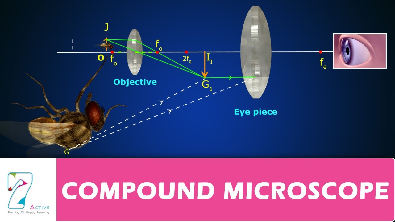
Compound microscope diagram. Diagram of a Compound Microscope - Biology Discussion Diagram of a Compound Microscope Article Shared by ADVERTISEMENTS: In this article we will discuss about:- 1. Essential Parts of Compound Microscope 2. Magnification of the Image of the Object by Compound Microscope 3. Resolution Power 4. Method for Studying Microbes 5. Measurement of the Size of Objects. Essential Parts of Compound Microscope: Compound Microscope: Diagram, Parts, Working & Magnification | AESL The figure below shows a compound microscope with its various parts. Working of compound microscope As shown in figure the lens near the object is called the objective which forms a real, inverted, magnified image of the object. This image serves as the object for the second lens. 16 Parts of a Compound Microscope: Diagrams and Video The 16 core parts of a compound microscope are: Head (Body) Arm Base Eyepiece Eyepiece tube Objective lenses Revolving Nosepiece (Turret) Rack stop Coarse adjustment knobs Fine adjustment knobs Stage Stage clips Aperture Illuminator Condenser Diaphragm Video: Parts of a compound Microscope with Diagram Explained Compound Microscope Principle, Parts, Diagram Definition, Application A compound microscope is a type of microscope that uses lenses and light to magnify objects. It consists of two main parts: the objective lens, which is located near the object being viewed, and the eyepiece, which is located near the viewer's eye.
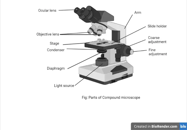
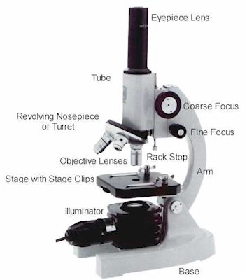
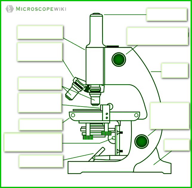
Compound Microscope Parts - Labeled Diagram and their Functions Labeled diagram of a compound microscope Major structural parts of a compound microscope There are three major structural parts of a compound microscope. The head includes the upper part of the microscope, which houses the most critical optical components, and the eyepiece tube of the microscope. Microscope - The compound microscope | Britannica compound microscope The microscope body tube separates the objective and the eyepiece and assures continuous alignment of the optics. It is a standardized length, anthropometrically related to the distance between the height of a bench or tabletop (on which the microscope stands) and the position of the seated observer's eyes. The compound microscope - how to draw ray diagrams - YouTube An animated presentation showing you how to draw ray diagrams (using simple lens rules) for a compound microscope. This shows how to determine the position a... Parts of the Microscope (Labeled Diagrams) - Simple and Compound Microscope Optical Parts of a Compound Microscope. Eyepiece lens or Ocular. Mirror. Objective Lenses. Scanning Objective Lens (4x) Low Power Objective (10x) High Power Objective Lens (40x) Oil Immersion Objective Lens (100x) Specialty Objective Lenses.
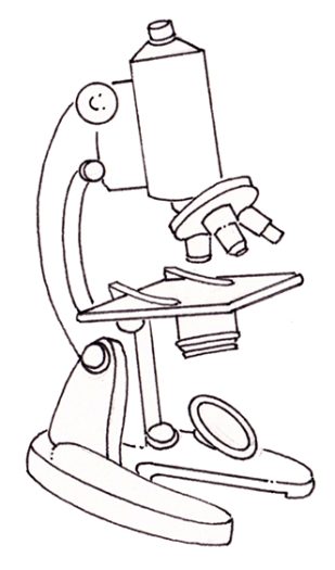
Compound Microscope - Diagram (Parts labelled), Principle and Uses Compound Microscope Parts (Labeled diagram) A compound microscope basically consists of optical and structural components. Within these two systems, there are multiple components within them and they are: Image : Labeled Diagram of compound microscope parts See: Labeled Diagram showing differences between compound and simple microscope parts Microscope | Types, Parts, History, Diagram, & Facts Single-lensed simple microscopes can magnify up to 300×—and are capable of revealing bacteria —while compound microscopes can magnify up to 2,000×. A simple microscope can resolve below 1 micrometre (μm; one millionth of a metre); a compound microscope can resolve down to about 0.2 μm. Labelled Diagram of Compound Microscope The below mentioned article provides a labelled diagram of compound microscope. Part # 1. The Stand: The stand is made up of a heavy foot which carries a curved inclinable limb or arm bearing the body tube. The foot is generally horse shoe-shaped structure (Fig. 2) which rests on table top or any other surface on which the microscope in kept. Compound Microscope Diagram Diagram | Quizlet Start studying Compound Microscope Diagram. Learn vocabulary, terms, and more with flashcards, games, and other study tools. ... The Compound Light Microscope Parts. 18 terms. Hollyster409. BIO 10 AP (part 3) 34 terms. AdamLister. Life Science: Cells Vocabulary. 12 terms. Julie_Clark647. What is Life? 13 terms.
Compound Microscope: Diagram, Parts, Working & Magnification ...
Parts of a microscope with functions and labeled diagram - Microbe Notes There are three structural parts of the microscope i.e. head, base, and arm. Head - This is also known as the body. It carries the optical parts in the upper part of the microscope. Base - It acts as microscopes support. It also carries microscopic illuminators.
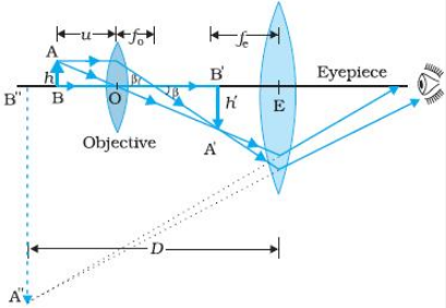
Draw a ray diagram to show the working of a compound ...
Compound Microscope: Definition, Diagram, Parts, Uses, Working Principle A compound microscope is defined as A microscope with a high resolution and uses two sets of lenses providing a 2-dimensional image of the sample. The term compound refers to the usage of more than one lens in the microscope. Also, the compound microscope is one of the types of optical microscopes.

Drawn and lable the diagram of compound microscope and ...
What is Compound Microscope? - Diagram, Function [updated] From the above Compound Microscope diagram, as you can observe, there are different types of parts like Eye Piece, Mirror, Base, Course Adjustment Knob, Nose Piece, Stage, Pillars, Base, Fine Adjustment Screw, Body Tube, Handle, Base, Pillars, Objective Lens, and many more.

Compound Microscope | Definition, Examples, Diagrams
Compound Microscope - Types, Parts, Diagram, Functions and Uses A compound microscope has the following: It comes with two or more convex lenses. One objective is used at a time. It produces 2-dimensional images. Its typical magnification is between 40x and 1000x. It is available in different configurations: monocular, binocular, and trinocular. (1, 2, 3, and 4)

Microscope Diagram Labeled, Unlabeled and Blank | Parts of a ...
Microscope Parts and Functions Here are the important compound microscope parts... Eyepiece: The lens the viewer looks through to see the specimen. The eyepiece usually contains a 10X or 15X power lens. Diopter Adjustment: Useful as a means to change focus on one eyepiece so as to correct for any difference in vision between your two eyes.

Draw a labelled ray diagram of compound microscope and derive ...
Microscopy: Intro to microscopes & how they work (article) - Khan Academy The fancier instruments that we typically think of as microscopes are compound microscopes, meaning that they have multiple lenses. Because of the way these lenses are arranged, they can bend light to produce a much more magnified image than that of a magnifying glass.
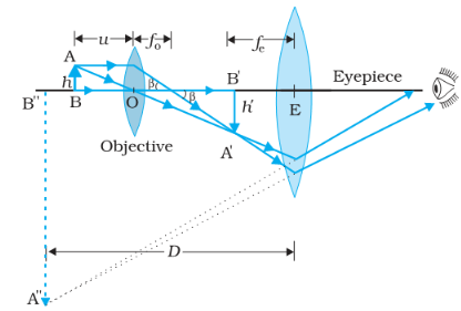
Draw a ray diagram to show the image formation by a compound ...
Compound Microscope Parts, Function, & Diagram - Study.com What are the parts of the compound microscope and their functions? There are many parts to the compound microscope. The eyepiece is the piece a person looks through. The objective lenses...
the compound microscope Diagram | Quizlet
Compound Microscope: Parts of Compound Microscope - BYJU'S The parts of the compound microscope can be categorized into: Mechanical parts; Optical parts (A) Mechanical Parts of a Compound Microscope. 1. Foot or base. It is a U-shaped structure and supports the entire weight of the compound microscope. 2. Pillar. It is a vertical projection. This stands by resting on the base and supports the stage. 3. Arm

How to draw compound of Microscope easily - step by step
Differences between Simple and Compound Microscope
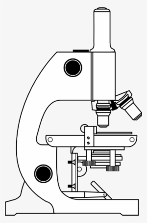
Small - Compound Microscope Black And White Transparent PNG ...

Compound microscope Diagram | Quizlet

4,029 Compound Microscope Images, Stock Photos & Vectors ...
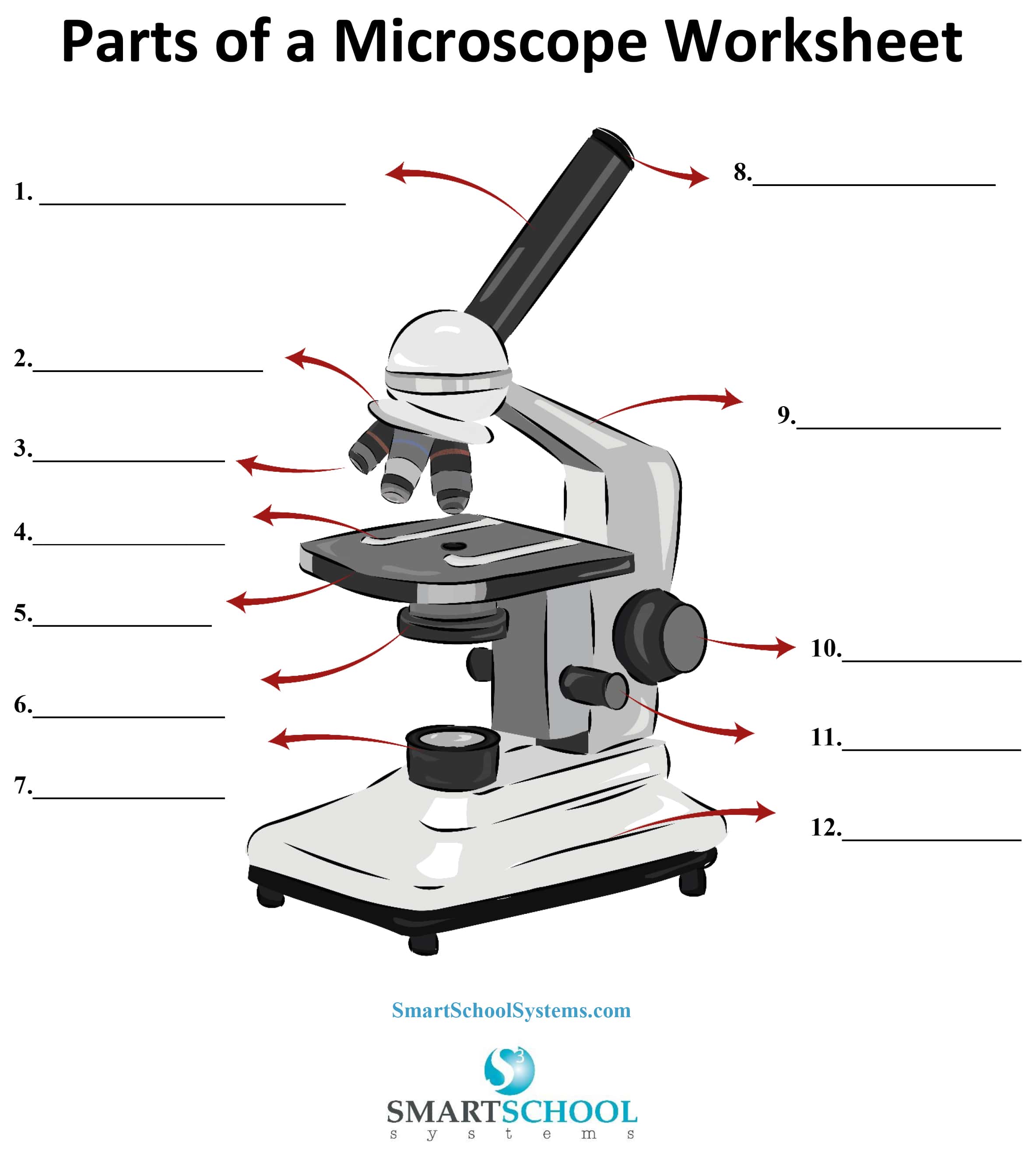
Parts of a Microscope - SmartSchool Systems

32: Ray diagram for compound microscope | Download Scientific ...

Simple doodles, Microscope parts, Microscope
Parts of a Microscope with Their Functions • Microbe Online
Microscope Parts & Specifications Labeled Diagram ...

HOw to draw light or compound microscope step by step / Microscope diagram

Draw a neat labelled diagram of a compound microscope. Derive ...

Compound Microscope Diagram | Quizlet

a Draw a ray diagram for the formation of image by a compound ...
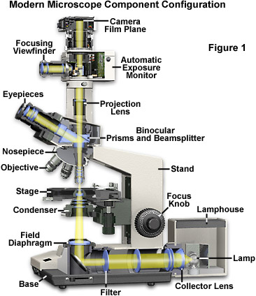
Molecular Expressions Microscopy Primer: Anatomy of the ...

Compound Microscope Parts – Labeled Diagram and their ...

Draw the ray diagram of image formation in case of compound ...

A Compound Microscope - Human Anatomy Figure by Monjurul ...
![Term 2] (a) Draw a ray diagram of compound microscope for ...](https://d1avenlh0i1xmr.cloudfront.net/6b555c3b-74ad-4391-b23d-c0b97c49ced1/ray-diagram---compound-microscope--final-image-at-d.jpg)
Term 2] (a) Draw a ray diagram of compound microscope for ...

Berkas:Microscope compound diagram.png - Wikipedia bahasa ...
COMPOUND MICROSCOPE

Microscope Diagram Labeled, Unlabeled and Blank | Parts of a ...
Difference Between Simple and Compound Microscope

Draw a Ray Diagram Showing the Image Formation by a Compound ...
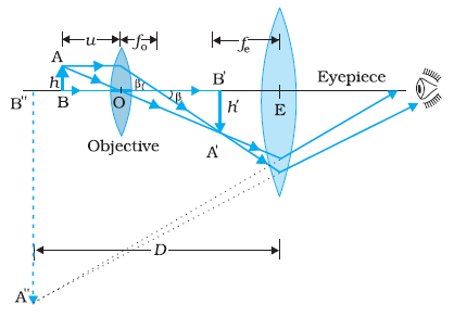
CBSE Class 12-science Answered
Compound microscope illustration - Lizzie Harper
Draw a labelled diagram of a compound microscope.
Simple Microscope - Diagram (Parts labelled), Principle ...
![What is Compound Microscope? - Diagram, Function [updated]](https://www.tutoroot.com/blog/wp-content/uploads/2022/09/Compound-microscope-1-300x273.png)
What is Compound Microscope? - Diagram, Function [updated]

Describe the structure of compound microscope with well ...

Lesson Explainer: Microscopy | Nagwa
Post a Comment for "39 compound microscope diagram"